WARNING:
JavaScript is turned OFF. None of the links on this concept map will
work until it is reactivated.
If you need help turning JavaScript On, click here.
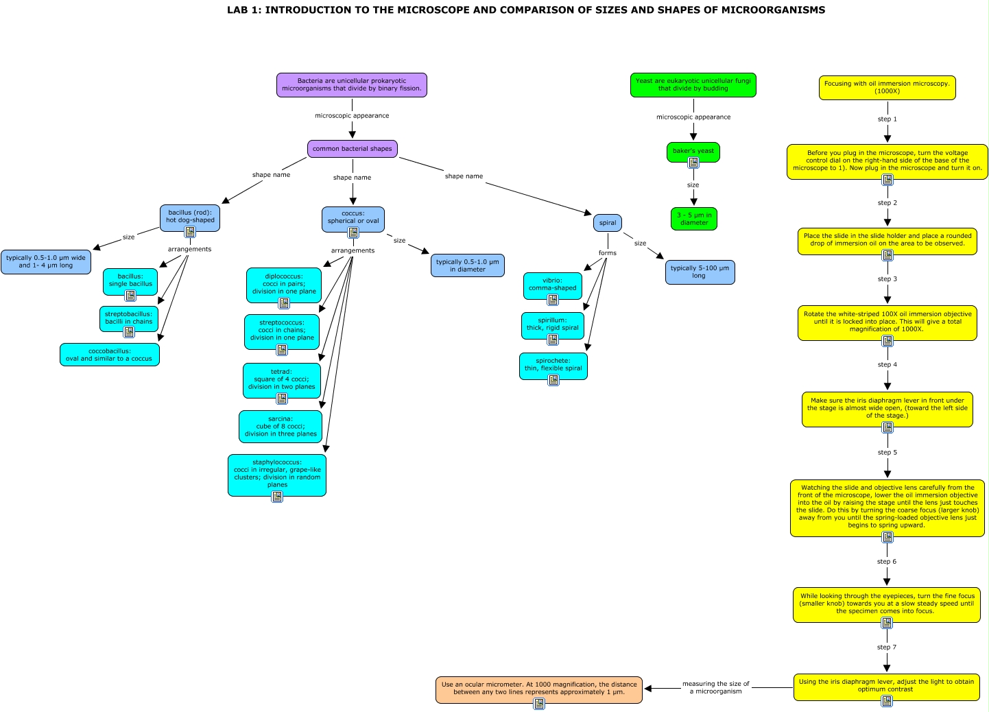
This Concept Map, created with IHMC CmapTools, has information related to: LAB 1- INTRODUCTION TO THE MICROSCOPE AND COMPARISON OF SIZES AND SHAPES OF MICROORGANISMS, coccus: spherical or oval arrangements staphylococcus: cocci in irregular, grape-like clusters; division in random planes, spiral forms spirochete: thin, flexible spiral, common bacterial shapes shape name coccus: spherical or oval, bacillus (rod): hot dog-shaped size typically 0.5-1.0 µm wide and 1- 4 µm long, Focusing with oil immersion microscopy. (1000X) step 1 Before you plug in the microscope, turn the voltage control dial on the right-hand side of the base of the microscope to 1). Now plug in the microscope and turn it on., Rotate the white-striped 100X oil immersion objective until it is locked into place. This will give a total magnification of 1000X. step 4 Make sure the iris diaphragm lever in front under the stage is almost wide open, (toward the left side of the stage.), Yeast are eukaryotic unicellular fungi that divide by budding microscopic appearance baker's yeast, common bacterial shapes shape name bacillus (rod): hot dog-shaped, common bacterial shapes shape name spiral, Place the slide in the slide holder and place a rounded drop of immersion oil on the area to be observed. step 3 Rotate the white-striped 100X oil immersion objective until it is locked into place. This will give a total magnification of 1000X., bacillus (rod): hot dog-shaped arrangements bacillus: single bacillus, Bacteria are unicellular prokaryotic microorganisms that divide by binary fission. microscopic appearance common bacterial shapes, coccus: spherical or oval arrangements diplococcus: cocci in pairs; division in one plane, coccus: spherical or oval size typically 0.5-1.0 µm in diameter, spiral forms vibrio: comma-shaped, spiral forms spirillum: thick, rigid spiral, baker's yeast size 3 - 5 µm in diameter, Using the iris diaphragm lever, adjust the light to obtain optimum contrast measuring the size of a microorganism Use an ocular micrometer. At 1000 magnification, the distance between any two lines represents approximately 1 µm., Watching the slide and objective lens carefully from the front of the microscope, lower the oil immersion objective into the oil by raising the stage until the lens just touches the slide. Do this by turning the coarse focus (larger knob) away from you until the spring-loaded objective lens just begins to spring upward. step 6 While looking through the eyepieces, turn the fine focus (smaller knob) towards you at a slow steady speed until the specimen comes into focus., While looking through the eyepieces, turn the fine focus (smaller knob) towards you at a slow steady speed until the specimen comes into focus. step 7 Using the iris diaphragm lever, adjust the light to obtain optimum contrast