WARNING:
JavaScript is turned OFF. None of the links on this concept map will
work until it is reactivated.
If you need help turning JavaScript On, click here.
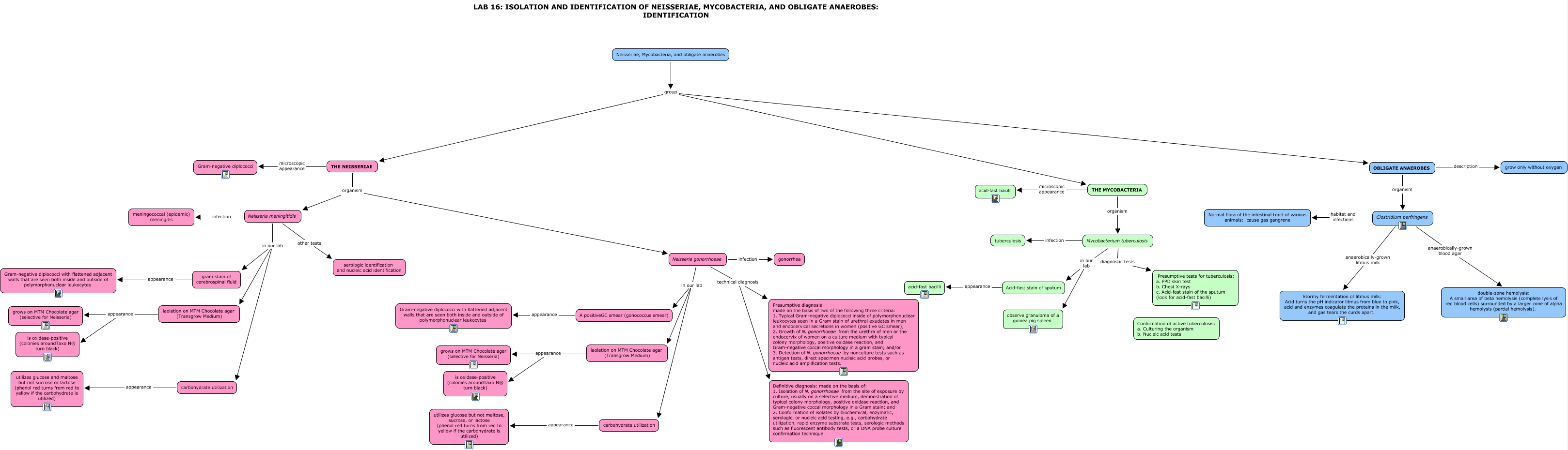
This Concept Map, created with IHMC CmapTools, has information related to: LAB 16 neisseria mycobacteria anaerobes identification, iaolation on MTM Chocolate agar (Transgrow Medium) appearance grows on MTM Chocolate agar (selective for Neisseria), gram stain of cerebrospinal fluid appearance Gram-negative diplococci with flattened adjacent walls that are seen both inside and outside of polymorphonuclear leukocytes, Clostridium perfringens anaerobically-grown blood agar double-zone hemolysis: A small area of beta hemolysis (complete lysis of red blood cells) surrounded by a larger zone of alpha hemolysis (partial hemolysis)., Neisseria gonorrhoeae in our lab A positiveGC smear (gonococcus smear), A positiveGC smear (gonococcus smear) appearance Gram-negative diplococci with flattened adjacent walls that are seen both inside and outside of polymorphonuclear leukocytes, Acid-fast stain of sputum appearance acid-fast bacilli, Clostridium perfringens anaerobically-grown litmus milk Stormy fermentation of litmus milk: Acid turns the pH indicator litmus from blue to pink, acid and enzymes coagulate the proteins in the milk, and gas tears the curds apart., Neisseria meningitidis infection meningococcal (epidemic) meningitis, carbohydrate utilization appearance utilizes glucose but not maltose, sucrose, or lactose (phenol red turns from red to yellow if the carbohydrate is utilized), OBLIGATE ANAEROBES organism Clostridium perfringens, iaolation on MTM Chocolate agar (Transgrow Medium) appearance is oxidase-positive (colonies aroundTaxo N® turn black), Mycobacterium tuberculosis infection tuberculosis, THE MYCOBACTERIA organism Mycobacterium tuberculosis, Neisseria gonorrhoeae technical diagnosis Presumptive diagnosis: made on the basis of two of the following three criteria: 1. Typical Gram-negative diplococci inside of polymorphonuclear leukocytes seen in a Gram stain of urethral exudates in men and endocervical secretions in women (positive GC smear); 2. Growth of N. gonorrhoeae from the urethra of men or the endocervix of women on a culture medium with typical colony morphology, positive oxidase reaction, and Gram-negative coccal morphology in a gram stain; and/or 3. Detection of N. gonorrhoeae by nonculture tests such as antigen tests, direct specimen nucleic acid probes, or nucleic acid amplification tests., Neisseria gonorrhoeae technical diagnosis Definitive diagnosis: made on the basis of: 1. Isolation of N. gonorrhoeae from the site of exposure by culture, usually on a selective medium, demonstration of typical colony morphology, positive oxidase reaction, and Gram-negative coccal morphology in a Gram stain; and 2. Conformation of isolates by biochemical, enzymatic, serologic, or nucleic acid testing, e.g., carbohydrate utilization, rapid enzyme substrate tests, serologic methods such as fluorescent antibody tests, or a DNA probe culture confirmation technique., Neisseriae, Mycobacteria, and obligate anaerobes group THE NEISSERIAE, Neisseriae, Mycobacteria, and obligate anaerobes group THE MYCOBACTERIA, Neisseria gonorrhoeae infection gonorrhea, THE NEISSERIAE organism Neisseria gonorrhoeae, iaolation on MTM Chocolate agar (Transgrow Medium) appearance is oxidase-positive (colonies aroundTaxo N® turn black)