WARNING:
JavaScript is turned OFF. None of the links on this concept map will
work until it is reactivated.
If you need help turning JavaScript On, click here.
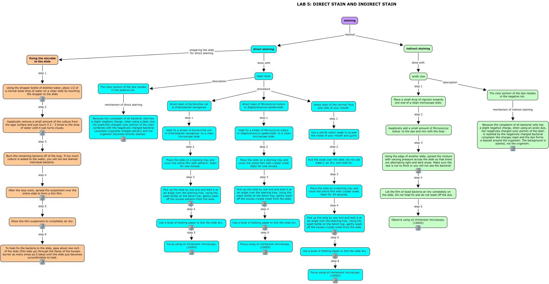
This Concept Map, created with IHMC CmapTools, has information related to: LAB 5 - DIRECT STAIN AND INDIRECT STAIN, direct staining done with basic dyes, basic dyes procedure direct stain of Micrococcus luteus or Staphylococcus epidermidis, acidic dye description The color portion of the dye resides in the negative ion., Place a small drop of nigrosin towards one end of a clean microscope slide. step 2 Aseptically add a small amount of Micrococcus luteus to the dye and mix with the loop., Let the film of dyed bacteria air dry completely on the slide. Do not heat fix and do not wash off the dye. step 5 Observe using oil immersion microscopy. (1000X), Heat fix a smear of Micrococcus luteus or Staphylococcus epidermidis to a clean microscope slide. step 2 Place the slide on a staining tray and cover the entire film with crystal violet. Stain for one minute., indirect staining done with acidic dye, Using the edge of another slide, spread the mixture with varying pressure across the slide so that there are alternating light and dark areas. Make sure the dye is not to thick or you will not see the bacteria! step 4 Let the film of dyed bacteria air dry completely on the slide. Do not heat fix and do not wash off the dye., basic dyes procedure direct stain of Escherichia coli or Enterobacter aerogenes, Allow this thin suspension to completely air dry. step 6 To heat-fix the bacteria to the slide, pass about one inch of the slide (film-side up) through the flame of the bunsen burner as many times as it takes until the slide just becomes uncomfortable to hold., Burn the remaining bacteria off of the loop. If too much culture is added to the water, you will not see stained individual bacteria. step 4 After the loop cools, spread the suspension over the entire slide to form a thin film., direct stain of Escherichia coli or Enterobacter aerogenes step 1 Heat fix a smear of Escherichia coli or Enterobacter aerogenes to a clean microscope slide., Use a book of blotting paper to blot the slide dry. step 5 Focus using oil immersion microscopy. (1000X), Place the slide on a staining tray and cover the entire film with safranin. Stain for one minute. step 3 Pick up the slide by one end and hold it at an angle over the staining tray. Using the wash bottle on the bench top, gently wash off the excess safranin from the slide., fixing the microbe to the slide step 1 Using the dropper bottle of distilled water, place 1/2 of a normal sized drop of water on a clean slide by touching the dropper to the slide, basic dyes procedure direct stain of the normal flora and cells of your mouth, The color portion of the dye resides in the positive ion. mechanism of direct staining Because the cytoplasm of all bacterial cells has a slight negative charge, when using a basic dye, the positively charged color portion of the stain combines with the negatively charged bacterial cytoplasm (opposite charges attract) and the organism becomes directly stained., Place the slide on a staining tray and cover the entire film with crystal violet. Stain for one minute. step 3 Pick up the slide by one end and hold it at an angle over the staining tray. Using the wash bottle on the bench top, gently wash off the excess crystal violet from the slide., Pick up the slide by one end and hold it at an angle over the staining tray. Using the wash bottle on the bench top, gently wash off the excess crystal violet from the slide. step 5 Use a book of blotting paper to blot the slide dry., Aseptically add a small amount of Micrococcus luteus to the dye and mix with the loop. step 3 Using the edge of another slide, spread the mixture with varying pressure across the slide so that there are alternating light and dark areas. Make sure the dye is not to thick or you will not see the bacteria!