Protozoans are unicellular eukaryotic microorganisms belonging to the Kingdom Protista. They reproduce asexually by fission (one cell splits into two), schizogony (multiple fission; the nucleus divides many times and the nuclei are separated into daughter cells), or budding (pinching off of a bud from a parent cell). Some protozoans also reproduce sexually by fusion of haploid sex cells called gametes.
The vegetative form (motile, feeding, reproducing form) of a protozoan is called a trophozoite. Under certain conditions, some protozoans produce a protective form called a cyst that enables them to survive harsh environments. Cysts allow some pathogens to survive outside their host. Favorable conditions in a new host result in excystation that once again produces a trophozoite.
The parasitic protozoans can be divided into groups based primarily on their means of locomotion.
1. The Sarcomastigophora (Amoeboflagellates)
a. The amoebas (subphylum Sarcodina) move by extending lobelike projections of their cytoplasm called pseudopodia. Food is obtained by phagocytosis.
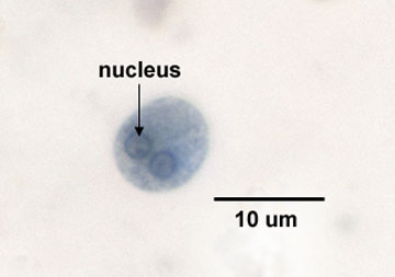
An important pathogen in this group is Entamoeba histolytica, the causative agent of amoebic dysentery. The organism is transmitted by the fecal-oral route. Cysts are excreted in the feces of an infected individual or carrier and ingested through fecally-contaminated food, water, objects, etc. After excystation, the trophozoites penetrate the walls of the large intestines causing ulceration and frequently causing the symptoms of dysentery. Involvement of the liver and other organs may occur if the protozoan invades the blood. The disease is diagnosed by microscopically looking for cysts of E. histolytica in a fecal smear (see Fig. 1).
Fig. 1: Cyst of Entamoeba histolytica in a fecal smear
Note one or more nuclei. Gary E. Kaiser, Ph.D.
Professor of Microbiology
The Community College of Baltimore County, Catonsville Campus
This work is licensed under a Creative Commons Attribution 3.0 Unported License
Acanthamoeba can infect the eye, blood, spinal cord, and brain and is transmitted by waterborne cysts picked up while swimming in contaminated water, crossing the mucous membranes.
b. The flagellates (subphylum Mastigophora) move by means of flagella. Some also have an undulating membrane.
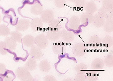
1. T. gambiense and T. rhodesiense cause the disease African sleeping sickness or African trypanosomiasis. They are transmitted to humans by the bite of an infected tsetse fly (a vector). The disease primarily involves the lymphatic and nervous systems of humans and is diagnosed by microscopically looking for Trypanosoma in the blood (see Fig. 2), in aspirated fluid from lymph nodes, or in spinal fluid.
Fig. 2: Trypanosoma gambiense in a blood smear
Note nucleus, flagellum, and undulating membrane. Gary E. Kaiser, Ph.D.
Professor of Microbiology
The Community College of Baltimore County, Catonsville Campus
This work is licensed under a Creative Commons Attribution 3.0 Unported License
T. cruzi causes South American sleeping sickness or Chagas' disease and is transmitted by infected Triatomid bugs (kissing bugs).
2. Giardia lamblia
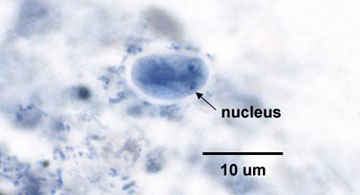
Giardia lamblia (G. intestinalis) causes a gastroenteritis-type of disease called giardiasis. Giardiasis is the most common protozoan intestinal disease in the U.S. and is transmitted by the fecal-oral route. Cysts of the organism are ingested through fecally-contaminated food, water, etc. Giardiasis is diagnosed by microscopically looking for cysts of G. lamblia in fecal smears (see Fig. 3).
Fig. 3: Giardia lamblia in a fecal smear
Note the two nuclei resembling "eyes." Gary E. Kaiser, Ph.D.
Professor of Microbiology
The Community College of Baltimore County, Catonsville Campus
This work is licensed under a Creative Commons Attribution 3.0 Unported License
- Scanning electron micrograph of Giardia; licensed for use, ASM MicrobeLibrary.
- Scanning electron micrograph of Giardia; courtesy of CDC.
3. Trichomonas vaginalis
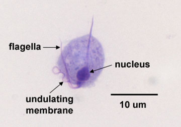
This protozoan causes genitourinary trichomoniasis. There are an estimated 2.5 million cases per year in the U.S. In females, it usually appears as vaginitis with itching and a white discharge. In males it is often asymptomatic but may cause urethritis. It is transmitted mainly by venereal contact and is diagnosed by microscopically looking for T. vaginalis trophozoites in vaginal discharge and urine (see Fig. 4).
Fig. 4: Trichomonas vaginalis in vaginal discharge
Note large nucleus, flagella, and undulating membrane. Gary E. Kaiser, Ph.D.
Professor of Microbiology
The Community College of Baltimore County, Catonsville Campus
This work is licensed under a Creative Commons Attribution 3.0 Unported License
2. The Ciliophora
This group of protozoans is characterized by a covering of cilia used for motility and direction of food particles into the mouth or cytosome. In the trophozoite, a large macronucleus, small micronucleus, cilia, and contractile vacuoles may be seen.
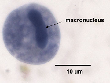
The only pathogen in this group is Balantidium coli, which causes a diarrhea-type infection called balantidiasis. The protozoan is transmitted to humans by the fecal-oral route and invades the large intestines causing ulceration. It is diagnosed by microscopically looking for B. coli in a fecal smear (Fig. 5).
Fig. 5: Balantidium coli in a fecal smear
Note large "dumbell-shaped" macronucleus (arrow). Gary E. Kaiser, Ph.D.
Professor of Microbiology
The Community College of Baltimore County, Catonsville Campus
This work is licensed under a Creative Commons Attribution 3.0 Unported License
3. The Apicomplexans
The sporozoa are not motile in their mature forms, reproduce both asexually and sexually, and often have complex life cycles for transmission from host to host. They possess a complex of organelles at their apex (apical complexes) that contain enzymes used in penetrating host tissues.
a. Toxoplasma gondii
This protozoan causes the disease toxoplasmosis. In adults, the disease is usually mild and resembles infectious mononucleosis. However, new-born infants who contracted toxoplasmosis in utero commonly have severe central nervous system damage. It also causes severe disease in immunosuppressed individuals such as people with AIDS. Domestic cats, that pick up the organism from eating infected rodents, may act as carriers of T. gondii, and their feces may contain oocysts of the protozoan. However, the organism may be found in practically every mammal. The disease is transmitted to humans by ingesting raw meat of an infected mammal or by inhaling or ingesting cysts of T. gondii from cat feces. Pregnant women should be especially careful to avoid raw meat and cat feces. The disease is diagnosed by serologic testing and by growing the organism in cell culture.
b. Plasmodium
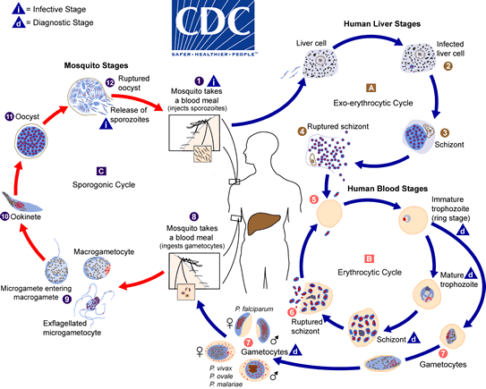
Four Plasmodium species, P. falciparum, P. malariae, P. ovale, and P. vivax cause malaria. The vector involved in the transmission of the disease from human to human or from animal to human is an infected female Anopheles mosquito.
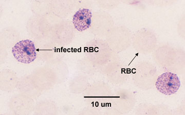
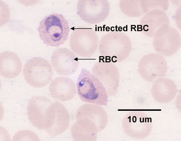
Asexual reproduction (or schizogony) of the Plasmodium occurs within liver cells and red blood cells of the infected human. With malaria caused by P. vivax and P. ovale, a dormant form or hypnozoite remains in the liver and may cause later relapses. The infected cells in which the organism is reproducing by schizogony are called schizonts. The sexual cycle (or sporogeny) occurs in the mosquito (see Fig. 6, the life cycle of Plasmodium). The typical recurring malarial fever is a result of the lysis of the infected red blood cells, causing release of merozoites and their metabolic by-products. Fever cycles of 24, 48, or 72 hours usually occur depending on the infecting species. Malaria is diagnosed by microscopically looking for the parasite within infected red blood cells (schizonts). See Fig. 7A and Fig 7B.
Fig. 6: Life cycle of Plasmodium, the protozoan that causes malaria
Fig. 7A: Plasmodium malariae infecting red blood cells
Fig. 7B: Plasmodium vivax infecting red blood cells
By Content Providers: CDC [Public domain].
Courtesy of the Centers for Disease Control and Prevention.Gary E. Kaiser, Ph.D.
Professor of Microbiology
The Community College of Baltimore County, Catonsville Campus
This work is licensed under a Creative Commons Attribution 3.0 Unported License
Gary E. Kaiser, Ph.D.
Professor of Microbiology
The Community College of Baltimore County, Catonsville Campus
This work is licensed under a Creative Commons Attribution 3.0 Unported License
c. Cryptosporidium
Cryptosporidium is an intracellular parasite that causes diarrhea, although in people who are immunosuppressed it can also cause respiratory and gallbladder infections. It is transmitted by the fecal-oral route.
Medscape articles on infections associated with organisms mentioned in this Lab Exercise. Registration to access this website is free.
B. PARASITIC HELMINTHS
Helminthology is the study of worms, or helminths. Over one billion people worldwide are infected with intestinal helminths alone. Helminths are multicellular, often macroscopic worms having both rudimentary organs and organ systems. We will look at three groups of pathogenic helminths: nematodes, cestodes, and trematodes.
1. The Nematodes (Roundworms)
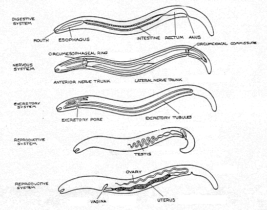
Nematodes are elongated, unsegmented, cylindrical worms having separate sexes. The various systems of the roundworms can be seen in Fig. 8, and a generalized drawing of a nematode is shown in Figs. 9A and 9B. We will look at several pathogenic nematodes.
Fig. 8: Organ Systems of Nematodes
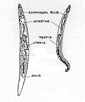
Fig. 9A: Morphology of Nematodes
Fig. 9B: Ascaris lumbricoides Gary E. Kaiser, Ph.D.
Professor of Microbiology
The Community College of Baltimore County, Catonsville Campus
This work is licensed under a Creative Commons Attribution 3.0 Unported License
Gary E. Kaiser, Ph.D.
Professor of Microbiology
The Community College of Baltimore County, Catonsville Campus
This work is licensed under a Creative Commons Attribution 3.0 Unported License
Gary E. Kaiser, Ph.D.
Professor of Microbiology
The Community College of Baltimore County, Catonsville Campus
This work is licensed under a Creative Commons Attribution 3.0 Unported License
a. Ascaris lumbricoides
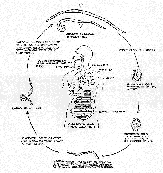
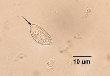
These worms range from 20-45 cm long and are 5 mm in diameter in the adult form, the female being larger than the male. The Ascaris life cycle is seen in Fig. 10. The disease is called ascariasis. This is the most common helminth in humans worldwide. In the U.S., it infects over 4,000,000 people. Humans become infected by ingesting water or food contaminated with feces that containsAscaris ova or from fingers contaminated with polluted soil. The disease is diagnosed by microscopically looking for Ascaris ova in a fecal smear (see Fig. 11). A similar roundworm, Toxocara, parasitizes dogs and cats. Visceral larva migrans is the migration of larvae of these worms in human tissues such as lung, liver, and brain, where they may cause tissue damage and allergic reactions.
Fig. 10: Life cycle of the nematode Ascaris lumbricoides
Fig. 11: Ova of Ascaris lumbricoides
Ova of Ascaris lumbricoides. Note "serrated edge" of ova (arrows). Gary E. Kaiser, Ph.D.
Professor of Microbiology
The Community College of Baltimore County, Catonsville Campus
This work is licensed under a Creative Commons Attribution 3.0 Unported License
Gary E. Kaiser, Ph.D.
Professor of Microbiology
The Community College of Baltimore County, Catonsville Campus
This work is licensed under a Creative Commons Attribution 3.0 Unported License
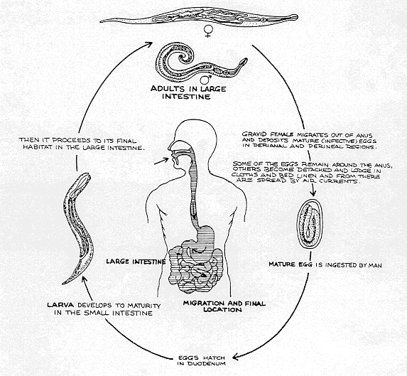
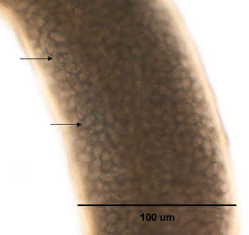
b. Enterobius vermicularis (pinworms)
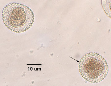
E. vermicularis is a small worm, the female being 8-13 mm long and 0.3-0.5 mm wide; the male being 2-5 mm long and 0.1 mm wide. The life cycle of Enterobiusis shown in Fig. 12. Pinworms are the most common helminth infection in the U.S. with as many as 50,000,000 people infected. Humans, frequently children, become infected by inhaling E. vermicularis ova or from transfer of ova to the mouth from fecally-contaminated fingers. The female worm migrates to the perianal region of the infected individual, releasing masses of ova and causing an itching sensation. Each pregnant worm produces between 4600 and 16,000 eggs (see Fig. 12A). The disease is diagnosed by applying tape to the perianal region and microscopically looking for pinworm ova that have stuck to the tape (see Fig. 13).
Fig. 12: Life cycle of the nematode Enterobius vermicularis (pinworm)
Fig 12A: Enterobius vermicularis (pinworm) filled with ova
Fig. 13: Ova of Enterobius vermicularis (pinworm)
; Central view of the nematode Enterobius vermicularis. Note that this female is filled with ova (arrows). Ova of Enterobius vermicularis. Note "smooth edge" of ova (arrows). Gary E. Kaiser, Ph.D.
Professor of Microbiology
The Community College of Baltimore County, Catonsville Campus
This work is licensed under a Creative Commons Attribution 3.0 Unported License
Gary E. Kaiser, Ph.D.
Professor of Microbiology
The Community College of Baltimore County, Catonsville Campus
This work is licensed under a Creative Commons Attribution 3.0 Unported License
Gary E. Kaiser, Ph.D.
Professor of Microbiology
The Community College of Baltimore County, Catonsville Campus
This work is licensed under a Creative Commons Attribution 3.0 Unported License
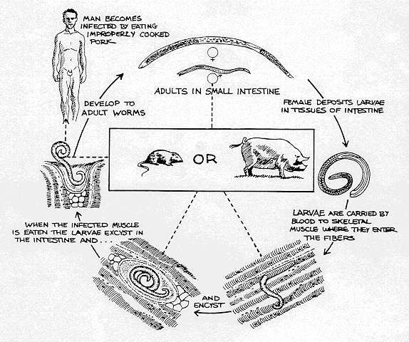
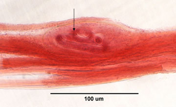
c. Trichinella spiralis
T. spiralis causes a disease called trichinosis. Humans become infected mainly by eating poorly cooked infected pork containing the encysted larva (1-2 mm long). The larvae excyst and develop into adult worms in the intestines. After mating, the female releases larvae which enter the blood and are distributed throughout the body where they become encysted in muscle tissue. The life cycle of Trichinella spiralis is seen in Fig. 14. The disease is diagnosed by serological tests and microscopic examination of biopsy specimens (see Fig. 15).
Fig. 14: Life cycle of the nematode Trichinella spiralis
Fig. 15: Trichinella spiralis Encysted in Muscle Tissue
Note spiraled larva of Trichinella (arrow) encysted in the muscle. Gary E. Kaiser, Ph.D.
Professor of Microbiology
The Community College of Baltimore County, Catonsville Campus
This work is licensed under a Creative Commons Attribution 3.0 Unported License
Gary E. Kaiser, Ph.D.
Professor of Microbiology
The Community College of Baltimore County, Catonsville Campus
This work is licensed under a Creative Commons Attribution 3.0 Unported License
2. The Cestodes (Tapeworms)
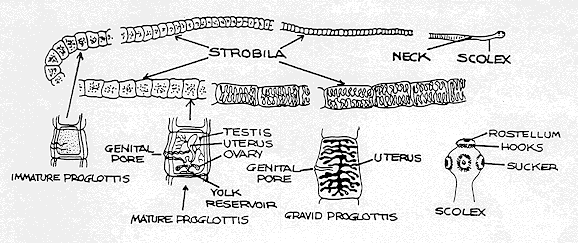
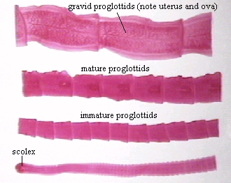
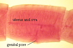
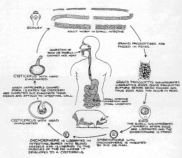
Tapeworms are flat segmented worms which are hermaphroditic (contain both male and female sexual organs). Adult tapeworms have several distinct regions (see Fig. 16A and 16B). The scolex (see Fig. 17) is a head-like structure with distinct suckers and possibly hooks used for attachment to the intestinal wall. Behind the scolex is a constricted neck region consisting of germinative tissue from which new segments, or proglottids, are formed. Finally, there is a long strobila or chain of proglottids of varying stages of maturity. Proglottids containing a uterus and thousands of ova are excreted in the feces (see Fig. 18). When ingested by intermediate hosts (such as cattle, pigs, and fish), the larva hatch from the ingested ova and migrate to muscle where they encyst as cysticerci. The life cycle of the pork tapeworm Taenia solium is shown in Fig. 19.
Fig. 16A: Tapeworm (Cestode) Morphology
Fig. 16B: The Tapeworm Taenia pisiformis (Cestode)
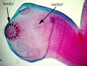
Fig. 17: Scolex of Taenia solium
Note the attachment structure or scolex, immature, mature, and gravid proglottids. Note hooks and suckers for attachment to intestinal wall. Gary E. Kaiser, Ph.D.
Professor of Microbiology
The Community College of Baltimore County, Catonsville Campus
This work is licensed under a Creative Commons Attribution 3.0 Unported License
Gary E. Kaiser, Ph.D.
Professor of Microbiology
The Community College of Baltimore County, Catonsville Campus
This work is licensed under a Creative Commons Attribution 3.0 Unported License
Gary E. Kaiser, Ph.D.
Professor of Microbiology
The Community College of Baltimore County, Catonsville Campus
This work is licensed under a Creative Commons Attribution 3.0 Unported License
Fig. 18: Gravid Proglottis of theTapeworm Taenia pisiformis (Cestode)
Fig. 19: Life cycle of the cestode Taenia solium (pork tapeworm)
The gravid proglottids are basically segments containing a uterus filled with ova and a genital pore for release of ova. Gary E. Kaiser, Ph.D.
Professor of Microbiology
The Community College of Baltimore County, Catonsville Campus
This work is licensed under a Creative Commons Attribution 3.0 Unported License
Gary E. Kaiser, Ph.D.
Professor of Microbiology
The Community College of Baltimore County, Catonsville Campus
This work is licensed under a Creative Commons Attribution 3.0 Unported License
Humans become infected with tapeworms by eating poorly cooked infected beef, pork, or fish containing cysticerci. Taenia saginata (See Fig. 20), the beef tapeworm often reaches 6 meters in length; Taenia solium, the pork tapeworm is normally 2-7 meters in length; and Diphyllobothrium latum, the fish tapeworm may reach 3-6 meters in length. These tapeworms are diagnosed by looking for proglottids and ova in the feces (See Fig. 21).
Fig. 20: The Tapeworm Taenia saginata (Cestode)
Fig. 21: Ovum of the Tapeworm Taenia saginata (Cestode)
The Tapeworm Taenia saginata (Cestode) [Public domain] Courtesy of the Centers for Disease Control and Prevention.
© Lynne Garcia, author. Licensed for use, ASM MicrobeLibrary.
When humans ingest tapeworm eggs instead of cysts, embryos are released, penetrate the intestinal wall, and enter the blood. The embryos migrate to various tissues (frequently the brain) and develop into cysticerci. Humans also act as intermediate hosts for Echinococcus granulosus found in dogs and cats. Larva hatch from ingested ova and migrate to the liver and lungs and form hydatid cysts.
3. The Trematodes (Flukes)
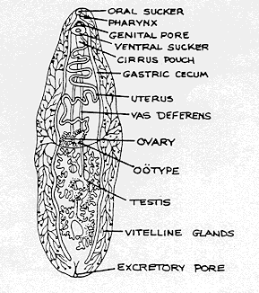
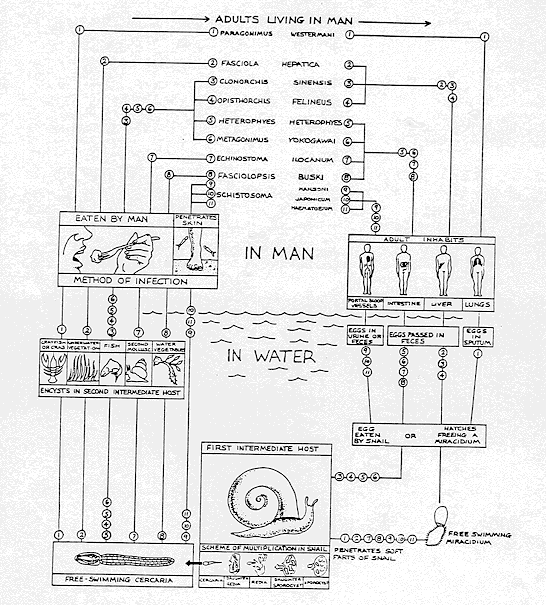
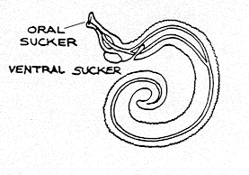
Flukes are unsegmented, flat, leaf-shaped worms having a variety of organ systems (see Fig. 22). Most flukes are hermaphroditic. They attach to the host by means of an oral sucker and a ventral sucker. Flukes, as adults, may infect either the portal blood vessels, intestines, liver, or lungs of humans and are named according to the tissue they infect. Humans become infected with liver flukes, lung flukes, and intestinal flukes by ingesting poorly cooked fish, crayfish, crabs, snails, or water vegetables infested with flukes. Blood flukes directly penetrate the skin.
In the life cycle of flukes (see Fig. 23), ova leave the body of the infected human or animal by means of feces, urine, or sputum (depending on the type of fluke). The ova enter water and infect the first intermediate host, certain species of water snails. A free swimming form of the fluke called the cercaria, then leaves the snail and infects second intermediate hosts (fish, crayfish, water vegetables, etc.) which are ingested by humans. The cercariae of the blood fluke Schistosoma (see Fig. 24) can directly penetrate the skin of humans and cause schistosomiasis, a major problem in Africa, South America, and Asia.
Fig. 22: Morphology of a Liver Fluke (Trematode)
Fig. 23: Life Cycle of Flukes (Trematodes)
Fig. 24: Morphology of a Blood Fluke (Trematode)
Gary E. Kaiser, Ph.D.
Professor of Microbiology
The Community College of Baltimore County, Catonsville Campus
This work is licensed under a Creative Commons Attribution 3.0 Unported License
Gary E. Kaiser, Ph.D.
Professor of Microbiology
The Community College of Baltimore County, Catonsville Campus
This work is licensed under a Creative Commons Attribution 3.0 Unported License
Gary E. Kaiser, Ph.D.
Professor of Microbiology
The Community College of Baltimore County, Catonsville Campus
This work is licensed under a Creative Commons Attribution 3.0 Unported License
Medscape articles on infections associated with organisms mentioned in this Lab Exercise. Registration to access this website is free.
1. Observe the prepared slides of the following parasitic protozoans and compare them with the indicated figures in this lab exercise.
a.The Sarcodina
Fecal smear containing cysts of Entamoeba histolytica (the cause of amoebic dysentery). Note that it contains several nuclei.
b. The Mastigophora
1. Blood smear containing Trypanosoma gambiense (the cause of African sleeping sickness). Note the nucleus, the undulating membrane, and the red blood cells in the background.
2. Fecal smear containing cysts and/or trophozoites of Giardia lamblia (the cause of giardiasis). Note the bilateral symmetry and macronuclei of the organism which look like "eyes."
3. Vaginal discharge containing Trichomonas vaginalis (the cause of genitourinary trichomoniasis). Note the bundle of flagella, the undulating membrane, and the nucleus.
c. The Ciliophora
Fecal smear containing Balantidium coli (the cause of balantidiasis). Note the large dumbbell-shaped macronucleus.
d. The Sporozoa
1. Sporozoites of Plasmodium from the salivary glands of an infected mosquito.
2. Blood smear containing red blood cells infected with merozoites of Plasmodium (the cause of malaria).
2. Observe the prepared slides of the following parasitic helminths and compare them with the indicated figures in this lab exercise.
a. The Nematodes (roundworms)
1. Fecal smear containing ova of Ascaris lumbricoides (the cause of ascariasis). Note the "bumpy" edge of the ova. The life cycle is shown in Fig. 10.
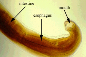
2. Ascaris lumbricoides larva. Note the organ systems.
3. Fecal smear containing ova of Enterobius vermicularis (pinworm). Note the "smooth" edge of the ova. The life cycle is shown in Fig. 12.
4. Enterobius vermicularis larva. Note the organ systems.
5. Muscle tissue containing encysted larvae of Trichinella spiralis (the cause of trichinosis). Note the spiral-shaped larva within the cyst. The life cycle is shown in Fig. 14.
b. The Cestodes (tapeworms)
1. Scolex of Taenia pisiformis (dog tapeworm). Note hooks and suckers.
2. Proglottids of the tapeworm Taenia pisiformis.
3. Gravid proglottid of Taenia pisiformis. Note the uterus and ova. Also see Fig. 16A.
c. The Trematodes (flukes)
1. Fasciola hepatica (liver fluke). Note the organ systems. See Fig. 22.
2. Schistosoma mansoni (blood fluke). Note the oral and ventral suckers. See Fig. 24.
3. Observe the preserved helminths.
PERFORMANCE OBJECTIVES FOR LAB 19
After completing this lab, the student will be able to perform the following objectives:
A. PARASITIC PROTOZOANS
DISCUSSION
1. Define the following: protozoan, trophozoite, cyst.
2. State how the following diseases may be transmitted to humans and briefly discuss how the diseases are diagnosed in the clinical laboratory:
a. amoebic dysentery
b. African sleeping sickness
c. giardiasis
d. genitourinary trichomoniasis
e. balantidiasis
f. malaria
g. toxoplasmosis
3. Describe the following in terms of the malarial life cycle: sporozoite, schizont, asexual cycle, sexual cycle.
4. State what causes the recurring fever of malaria.
RESULTS
1. Recognize the following organisms when seen through a microscope and state what disease they are associated with:
a. Entamoeba histolytica cysts in a fecal smear
b. Trypanosoma gambiense in a blood smear
c. Giardia lamblia cysts in a fecal smear
d. Trichomonas vaginalis in vaginal discharge
e. Balantidium coli in a fecal smear
f. Plasmodium merozoites in infected red blood cells
B. PARASITIC HELMINTHS
DISCUSSION
1. Define the following: helminth, ova, hermaphroditic.
2. List the three classes of parasitic helminths and state the common name for each class.
3. State how the following diseases may be transmitted to humans and state how each disease may be diagnosed in the clinical laboratory:
a. ascariasis
b. pinworms
c. trichinosis
d. tapeworms
e. flukes
4. Compare protozoans and helminths in terms of their size and structural complexity.
RESULTS
1. Recognize the following organisms or structures when seen through a microscope:
a. roundworms
b.Ascaris ova
c. pinworm ova
d. Trichinella in muscle tissue
e. tapeworms
f. scolex of a tapeworm
g. ova and uterus in a gravid proglottid of a tapeworm
h. flukes
SELF-QUIZ

Microbiology Laboratory Manual by Gary E. Kaiser, PhD, Professor of Microbiology
is licensed under a Creative Commons Attribution 4.0 International License.
Last updated: September, 2023