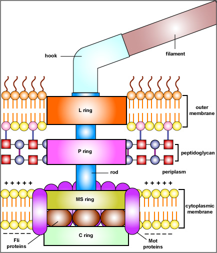
Fig. 12: Structure of a Bacterial Flagellum

The filament of the bacterial flagellum is connected to a hook which, in turn, is attached to a rod. The basal body of the flagellum consists
of a rod and a series of rings that anchor the flagellum to the cell wall
and the cytoplasmic membrane. In gram-negative bacteria, the L ring anchors the flagellum to the lipopolysaccharide layer of the outer membrane while the P ring anchors the flagellum to the peptidoglycan portion of the cell wall. The MS ring is located in the cytoplasmic membrane and the C ring in the cytoplasm. The Mot proteins surround the MS and C rings of the motor and function to generate torque for rotation of the flagellum. Energy for rotation comes from proton motive force. Protons moving through the Mot proteins drives rotation. The Fli proteins act as the motor switch to trigger either clockwise or counterclockwise rotation of the flagellum and to possibly disengage the rod in order to stop motility.
Illustration of the Structure of a Bacterial Flagellum .jpg by Gary E. Kaiser, Ph.D.
Professor of Microbiology,
The Community College of Baltimore County, Catonsville Campus
This work is licensed under a Creative Commons Attribution 4.0 International License.
Based on a work at https://cwoer.ccbcmd.edu/science/microbiology/index_gos.html.

Last updated: Feb., 2021
Please send comments and inquiries to Dr.
Gary Kaiser
