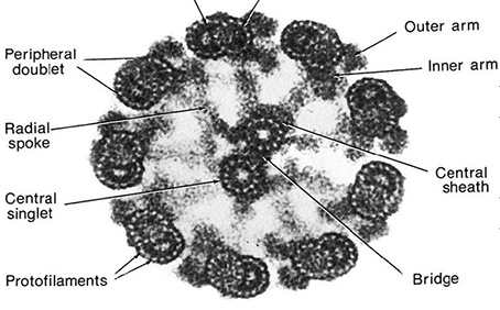
Fig. 44: Transmission Electron Micrograph of a Cilium

This thin section electron micrograph shows a cross section of a cilium from a rat tracheal epithelial cell. Note the 2 X 9 + 2 arrangement of microtubules and the dynein arms.
Fig. 44: Transmission Electron Micrograph of a Cilium

This thin section electron micrograph shows a cross section of a cilium from a rat tracheal epithelial cell. Note the 2 X 9 + 2 arrangement of microtubules and the dynein arms.
Don W. Fawcett (2011) CIL:11626, Rattus sp., tracheal epithelial cell. CIL. Dataset. https://doi.org/doi:10.7295/W9CIL11626

Attribution Non-Commercial; No Derivatives:This image is licensed under a Creative Commons Attribution, Non-Commercial, No Derivatives License. View License Deed View Legal Code