Fig. 5: Trichomonas vaginalis
in Vaginal Discharge
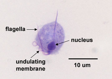
Note large nucleus, flagella, and
undulating membrane.
Photomicrograph of Trichomonas vaginalis .jpg by Gary E. Kaiser, Ph.D.
Professor of Microbiology,
The Community College of Baltimore County, Catonsville Campus
This work is licensed under a Creative Commons Attribution 4.0 International License.
Based on a work at https://cwoer.ccbcmd.edu/science/microbiology/index_gos.html.

Last updated: Feb., 2018
Please send comments and inquiries to Dr.
Gary Kaiser