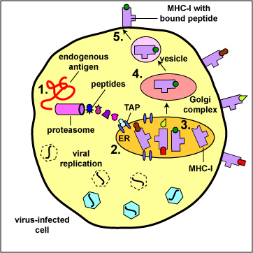
The body marks infected cells and tumor cells for destruction by placing peptide epitopes from these endogenous antigens on their surface by way of MHC-I molecules. Cytotoxic T-lymphocytes (CTLs) are then able to recognize peptide/MHC-I complexes by means of their T-cell receptors (TCRs) and CD8 molecules and kill the cells to which they bind.
1. During viral replication within the host cell, endogenous antigens, such as viral proteins, pass through proteasomes where they are degraded into a series of peptides.
2. The peptides are transported into the rough endoplasmic reticulum (ER) by a transporter protein called TAP.
3. The peptides then bind to the grooves of newly synthesized MHC-I molecules.
4. The endoplasmic reticulum transports the MHC-I molecules with bound peptides to the Golgi complex.
5. The Golgi complex, in turn, transports the MHC-I/peptide complexes by way of an exocytic vesicle to the cytoplasmic membrane where they become anchored. Here, the peptide and MHC-I/peptide complexes can be recognized by CTLs by way of TCRs and CD8 molecules having a complementary shape.
Last updated: Feb., 2021
Please send comments and inquiries to Dr.
Gary Kaiser