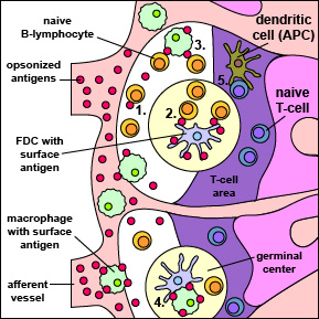
Fig. 3B: A B-lymphocyte Recognizing
Antigens on the Surface of Specialized
Macrophages and Follicular Dendritic Cells (FDCs) Located in Lymph Nodes

Opsonized antigens (those coated with C3b and Ced from the complement pathways) enter a lymph node through afferent lymphoid vessels. These opsonized antigens bind to and remain on the surface of specialized macrophages and follicular dendritic cells (FDCs). In addition, macrophages can transfer antigens to FDCs (see 4. above). Using their B-cell receptor (BCR), naive B-lymphocytes are able to recognize antigens directly (see 1. above), or more commonly, on the surface of FDCs (see 2. above), or on the surface of macrophages (see 3. above) in the germinal centers and lymphoid follicles of the lymph node. Meanwhile, naive T-lymphocytes are being activated by antigen-presenting dendritic cells in the T-cell areas of the lymph node (see 5. above). T4-effector cells and activated B-lymphcytes then interact with one another at the interface between the geminal centers and the T-cell areas.
Illustration of A B-lymphocyte Recognizing Antigens on the Surface of Specialized Macrophages and Follicular Dendritic Cells (FDCs) Located in Lymph Nodes .jpg by Gary E. Kaiser, Ph.D.
Professor of Microbiology,
The Community College of Baltimore County, Catonsville Campus
This work is licensed under a Creative Commons Attribution 4.0 International License.
Based on a work at https://cwoer.ccbcmd.edu/science/microbiology/index_gos.html.

Last updated: August, 2019
Please send comments and inquiries to Dr.
Gary Kaiser
