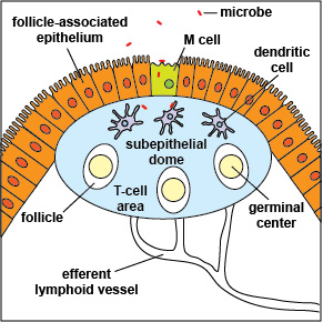
Peyer's patches are part of the mucosa-associated lymphoid tissue (MALT) in the small intestines. Microbes and antigens enter through specialized epithelial cells called microfold (M) cells. Changing populations of naive B-lymphocytes and naive T-lymphocytes enter the Peyer's patch via blood vessels with B-lymphocytes entering the follicles and germinal centers and T-lymphocytes entering the T-cell area. Dendritic cells engulf and process antigens and present them by way of MHC molecules to the TCRs of naive T-lymphocytes.
Last updated: August, 2019
Please send comments and inquiries to Dr.
Gary Kaiser
