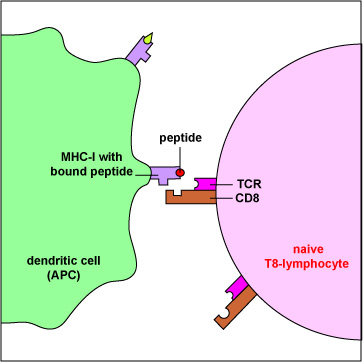
Antigen-presenting dendritic cells produce both MHC-I and MHC-II molecules. These dendritic cells can phagocytose infected cells and tumor cells, place them in phagosomes, and degrade them with lysosomes. During this process, some of the proteins escape from the phagosome into the surrounding cytosol. Here they can be degraded into peptides by proteasomes, bound to MHC-I molecules, and placed on the surface of the dendritic cell. Now the peptide/MHC-I complexes can be recognized by a naive T8-lymphocyte having a complementary shaped T-cell receptor (TCR) and CD8 molecule. This activates the naive T8-lymphocyte enabling it to eventually proliferate and differentiate into cytotoxic T-lymphocytes (CTLs).
Last updated: August, 2019
Please send comments and inquiries to Dr.
Gary Kaiser