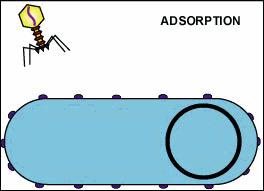
Attachment sites on the bacteriophage adsorb to receptor sites on the host bacterium (see Fig. 1). Most bacteriophages adsorb to the bacterial cell wall, although some are able to adsorb to flagella or pili. Specific strains of bacteriophages can only adsorb to specific strain of host bacteria. This is known as viral specificity (def).
by Gary E. Kaiser, Ph.D.
Professor of Microbiology, The Community College of Baltimore County, Catonsville Campus
This work is licensed under a Creative Commons Attribution 4.0 International License.
Based on a work The Grapes of Staph at https://cwoer.ccbcmd.edu/science/microbiology/index_gos.html.Last updated: August, 2019
Please send comments and inquiries to Dr. Gary Kaiser

In the case of bacteriophages that adsorb to the bacterial cell wall, a bacteriophage enzyme "drills" a hole in the bacterial wall and the bacteriophage injects its genome into the bacterial cytoplasm (see Fig. 2). Some bacteriophages accomplish this by contracting a sheath which drives a hollow tube into the bacterium. This begins the eclipse period. The genomes of bacteriophages which adsorb to flagella or pili enter through these hollow organelles. In either case, only the phage genome enters the bacterium so there is no uncoating stage.
by Gary E. Kaiser, Ph.D.
Professor of Microbiology, The Community College of Baltimore County, Catonsville Campus
This work is licensed under a Creative Commons Attribution 4.0 International License.
Based on a work The Grapes of Staph at https://cwoer.ccbcmd.edu/science/microbiology/index_gos.html.Last updated: August, 2019
Please send comments and inquiries to Dr. Gary Kaiser
Enzymes coded by the bacteriophage genome shut down the bacterium's macromolecular (protein, RNA, DNA) synthesis. The bacteriophage replicates its genome and uses the bacterium's metabolic machinery to synthesize bacteriophage enzymes and bacteriophage structural components (see Fig. 3 and Fig. 4).
by Gary E. Kaiser, Ph.D.
Professor of Microbiology, The Community College of Baltimore County, Catonsville Campus
This work is licensed under a Creative Commons Attribution 4.0 International License.
Based on a work The Grapes of Staph at https://cwoer.ccbcmd.edu/science/microbiology/index_gos.html.Last updated: August, 2019
Please send comments and inquiries to Dr. Gary Kaiser
4. Maturation
The phage parts assemble around the genomes (see Fig. 5).
by Gary E. Kaiser, Ph.D.
Professor of Microbiology, The Community College of Baltimore County, Catonsville Campus
This work is licensed under a Creative Commons Attribution 4.0 International License.
Based on a work The Grapes of Staph at https://cwoer.ccbcmd.edu/science/microbiology/index_gos.html.Last updated: August, 2019
Please send comments and inquiries to Dr. Gary Kaiser
Usually, a bacteriophage-coded lysozyme breaks down the bacterial peptidoglycan causing osmotic lysis and release of the intact bacteriophages (see Fig. 6).
by Gary E. Kaiser, Ph.D.
Professor of Microbiology, The Community College of Baltimore County, Catonsville Campus
This work is licensed under a Creative Commons Attribution 4.0 International License.
Based on a work The Grapes of Staph at https://cwoer.ccbcmd.edu/science/microbiology/index_gos.html.Last updated: August, 2019
Please send comments and inquiries to Dr. Gary Kaiser
YouTube movie showing the lysis of Escherichia coli by coliphage T4.
Courtesy of Jim Sullivan, Cells Alive
From 50 to 200 bacteriophages may be produced per infected bacterium.
by Gary E. Kaiser, Ph.D.
Professor of Microbiology, The Community College of Baltimore County, Catonsville Campus
This work is licensed under a Creative Commons Attribution 4.0 International License.
Based on a work The Grapes of Staph at https://cwoer.ccbcmd.edu/science/microbiology/index_gos.html.Last updated: August, 2019
Please send comments and inquiries to Dr. Gary Kaiser